Definition
Radiology is a branch of medicine using radiation for the diagnosis and treatment of disease. Radiology originally involved the use of X rays in the diagnosis of disease and the use of X rays, gamma rays, and other forms of ionizing radiation in the treatment of disease. In more recent years radiology has come also to embrace diagnosis by a method of organ scanning with the use of radioactive isotopes and also with nonionizing radiation, such as ultrasound waves and nuclear magnetic resonance. Similarly, the scope of radiotherapy has extended to include, in the treatment of cancer, such agents as hormones and chemotherapeutic drugs.
History
X rays were discovered by Wilhelm Conrad Röntgen, a German professor of physics, in his laboratory in the University of Würzburg on Nov. 8, 1895. Early on, in radiodiagnosis, use was made of three of the properties of X rays--their ability to penetrate the tissues, their photographic effect, and their ability to cause certain substances to fluoresce. In penetrating the tissues, the radiation is absorbed differentially, depending on the densities of the tissues being penetrated. The radiation emerging from tissues thus produces on a photographic film or a fluorescent screen an image of the structures of differing densities within the body. The limiting factor in this method of diagnosis is the similarity between the densities of adjacent soft tissues within the body, with a resultant failure to produce a notable contrast between the images of adjacent structures or organs.
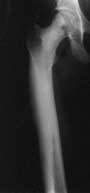 During the first two decades following their discovery, X rays were used largely for the diagnosis and control of treatment of fractures and for the localization of foreign bodies, such as bullets, during World War I. The physicians using these methods introduced artificial contrast agents, such as a paste consisting of barium sulfate, which is inert and nontoxic when taken by mouth. When a contrast agent is taken by mouth or introduced by enema, the various parts of the alimentary tract can be demonstrated and examined. Refinements of this technique continue to the present day, and radiological examination of the alimentary tract is an elegant and precise aid to diagnosis. Eventually a number of other contrast media were produced that could be injected into blood vessels. The media could thus be used either to demonstrate those vessels (whether arteries or veins) or, after their selective concentration and excretion by the kidneys, to show the urinary tract.
Within the first few months after Röntgen's discovery, attempts were made to produce films of moving objects; thus, it was soon realized that radiology might be able to depict function and so demonstrate dynamic physiological functions rather than just static anatomy. Technical difficulties and the hazards of a high dose of radiation to the patient prevented the proper development of this technique.
Image intensifier
In the 1950s an electronic method was devised to intensify the image, the so-called image intensifier, which made possible the overcoming of the technical difficulties, and cineradiography became routine. During the whole period of the development of radiology, photographic techniques were also continually being improved. Single-coated photographic plates were used at first, and then double-coated photographic films; photographic emulsions have now been developed to such a point that high speed can be provided with good definition and little intrusion of photographic grain into the image. Similarly, processing methods have been improved; automatic processors now can deliver a fully processed dry film in 90 seconds.
CAT scanning
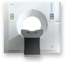 A new form of X-ray imaging, computerized axial tomography (CAT scanning), was devised by Godfrey Hounsfield of Great Britain and Allan Cormack of the United States during the 1970s. This method measures the attenuation of X-rays entering the body from many different angles. From these measurements a computer reconstructs the organ under study in a series of cross sections or planes. The technique allows soft tissues such as the liver and kidney to be clearly differentiated in the images reconstructed by the computer. This procedure adds enormously to the diagnostic information that can be provided by conventional X rays. CAT scanners are now in use in many large hospitals and medical centres throughout the world.
Nuclear magnetic resonance
A still more recently developed technique is nuclear magnetic resonance (NMR) scanning (also called magnetic resonance imaging, or MRI), in which radio waves are beamed into an individual who is subjected to a powerful magnetic field. Different atoms in the body absorb radio waves at different frequencies under the influence of the magnetic field. The way in which absorption takes place is measured and used by a computer to construct images of internal structures.
PET scanning
Another recent technique is positron emission tomography, or PET scanning, which involves the emission of particles of antimatter by compounds injected into the body being scanned. These particles, positrons, are neutralized by their opposites, electrons, and energy is released in the form of radiation as matter and antimatter annihilate each other. Detectors arranged around the body pick up the energy released and use it to follow the movements of the injected compound and its metabolism.
These relatively new radiological techniques provide much safer means of examining internal body structures. They also yield precise and clear images for the physician and diminish the margin of error in therapeutic measures.
Radiotherapy
In radiotherapy, use is made of the biological effects of ionizing radiations. The early workers noted that large doses of radiation would cause, after some delay, reddening of the skin, which might lead to blistering and ulceration. Even small repeated doses, if occurring often enough, might produce serious skin lesions. It was argued, then, that a phenomenon producing such damage to normal tissues might be directed toward abnormal and undesirable tissues, such as cancer. Research into the fundamental nature of the biological action of radiation continues to the present day, and a new type of scientist, the radiobiologist, has emerged. About the same time as the uses of X rays were first being applied to medicine, radium was discovered, and also the importance of the time factor as a modifier of the reaction of tissue to radiation was established. Thus was born the art of radiotherapy, at first based entirely on an empirical approach. It was soon noted that ionizing radiations also have the effect of alleviating pain, and so in the period of development of this form of treatment it was used rather extensively in the treatment of painful forms of arthritis, swellings of the salivary glands, herpes zoster or shingles, overgrowth of adenoids in children, and several other benign conditions. As knowledge of the possible harmful effects of radiation has grown, many of these applications have been discarded, except in special circumstances and under strict supervision.
The vast bulk of the practice of radiotherapy has to do with cancer, and it is here that the great advances have been made. The first step was to establish a unit of measurement. Since the early days physicians practicing treatment with ionizing radiations have worked in close collaboration with physicists, and much of the fundamental research has been undertaken by radiation physicists working alongside their medical colleagues. The establishment of an internationally accepted unit of measurement at Stockholm in 1925, the roentgen unit, enabled physicists to undertake similar calibration in different centres and provided a means of devising a system of dosimetry.
Links