
An electroencephalogram (EEG) is a test that looks at the function of the brain. The brain works by a series of nerve impulses, which cause electrical signals. These signals (also called brainwaves) can be recorded through the scalp.
Measuring EEG electrical activity
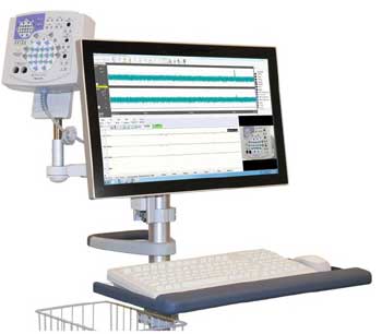 EEG is a method to record the electrical activity on the scalp that has been shown to represent the macroscopic activity of the surface layer of the brain underneath. It is typically non-invasive, with the electrodes placed along the scalp. Electrocorticography, involving invasive electrodes, is sometimes called "intracranial EEG".
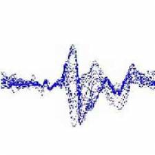
EEG measures voltage fluctuations resulting from ionic current within the neurons of the brain. Clinically, EEG refers to the recording of the brain's spontaneous electrical activity over a period of time, as recorded from multiple electrodes placed on the scalp. Diagnostic applications generally focus either on event-related potentials or on the spectral content of EEG. The former investigates potential fluctuations time locked to an event, such as 'stimulus onset' or 'button press'. The latter analyses the type of neural oscillations (popularly called "brain waves") that can be observed in EEG signals in the frequency domain.
EEG is most often used to diagnose epilepsy, which causes abnormalities in EEG readings. It is also used to diagnose sleep disorders, depth of anaesthesia, coma, encephalopathies, and brain death. EEG used to be a first-line method of diagnosis for tumours, stroke and other focal brain disorders, but this use has decreased with the advent of high-resolution anatomical imaging techniques such as magnetic resonance imaging (MRI) and computed tomography (CT). Despite limited spatial resolution, EEG continues to be a valuable tool for research and diagnosis. It is one of the few mobile techniques available and offers millisecond-range temporal resolution which is not possible with CT, PET or MRI.
Derivatives of the EEG technique include evoked potentials (EP), which involves averaging the EEG activity time-locked to the presentation of a stimulus of some sort (visual, somatosensory, or auditory). Event-related potentials (ERPs) refer to averaged EEG responses that are time-locked to more complex processing of stimuli; this technique is used in cognitive science, cognitive psychology, and psychophysiological research.
Why carry out a sleep study?
Common reasons for a sleep study include:
Excessive snoring
Sleep apnoea (periods where the breath stops)
Daytime sleepiness
Insomnia (inability to sleep)
Narcolepsy (sudden onset of sleep)
Restless legs syndrome (condition causing uncomfortable leg sensations)
Nightmares during nondream stages of sleep (sleep terrors), sleep walking or talking, and rapid eye movement disorders are less common conditions that may also require a sleep study.
How is a sleep EEG used?
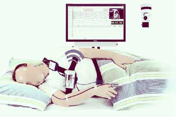
Recording the EEG during drowsiness and sleep can be useful to identify brain activity which may not be present while awake. It can be carried out on patients of all ages and abilities, making it a good way to get an overall view of the function of the brain. It is helpful as part of general investigations and more specific problems, such as blackouts and seizures.
The stages of sleep range from light to deep. Each stage has characteristics that can be measured. A sleep study is a number of tests done at the same time during sleep. The tests measure specific sleep characteristics and help to diagnose sleep disorders. A sleep study may also be called polysomnogram.
The basic recordings done during a sleep study may include:
Electroencephalography (EEG) - This measures brain wave activity.
Electrooculogram (EOG) - This measures eye movement.
Electromyography (EMG) - This measures muscle movement.
Other recordings - An electrocardiogram (ECG) is used to record electrical activity of the heart.
 Sleep studies take place in a sleep lab during your normal sleeping hours. The goal is to record brain and body activity that happens during sleep so that sleep disorders can be diagnosed and treated.
Video monitoring - Simultaneous video monitoring of the patient during the EEG recording is becoming more popular. It allows the physician to closely correlate EEG waveforms with the patients activity and may help produce a more accurate diagnosis.
Video recorders and cameras can be connected to an EEG machine using a time code generator. This records an accurate time signal onto the videotape. When the video and EEG are reviewed together the two signals are accurately synchronised together. Video monitoring is always used for Long Term Monitoring recordings as the patient is unattended. The patient may also have an event button connected to the EEG machine so that times when the patient thought they were having an epileptic attack can be easily identified.
During a sleep study, the following may be measured:
Eye movement - The number of eye movements and their frequency or speed.
Brain activity - The electrical currents of the brain.
Limb movement - The number and intensity of movements.
Breathing patterns - The number and depth of respirations.
Heart rhythm - The electrical activity of the heart.
Oxygen saturation - The percentage of oxygen in the blood.
Acid/base balance of the stomach - The amount of acid secreted during sleep.
Sleep latency - The time it takes to fall asleep.
Sleep duration - The period of time a person stays asleep.
Sleep efficiency - The ratio of the total time asleep to the total time in bed.
In addition, these tests may be done:
Multiple sleep latency tests (MSLT) measure how long it takes to fall asleep.
Multiple wake tests (MWT) measure whether you can stay awake during specified times.
Doctors trained in sleep medicine evaluate test results to treat sleep issues. A trained sleep technician will be with you in the sleep lab during the testing period.
Sources:
https://www.nhs.uk/conditions/electroencephalogram/
https://www.hopkinsmedicine.org/health/treatment-tests-and-therapies/sleep-study
https://en.wikipedia.org/wiki/Electroencephalography
https://eu.nihonkohden.com/sites/default/files/styles/product_image_large/public/nihonkohden-neurofax_eeg_1200-side-l.jpg
https://www.ebme.co.uk/articles/clinical-engineering/introduction-to-eeg


